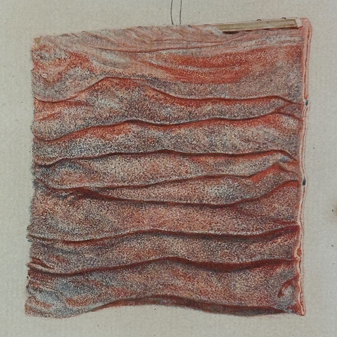
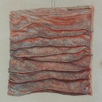
Icon tunicæ villosæ intestini duodeni
| Macro-category | Rare Books and Manuscripts |
| Subcategory | Medicine |
| Author | BLEULAND, J. |
| Place of printing | Trajecti ad Rhenum |
| Publisher / Maker | B. Wild et J. Altheer |
| Year | 1789 |
a new era for Anatomical Pathology:
The rise of coloured illustrations
First edition of Bleuland’s histological investigation of duodenal tissue
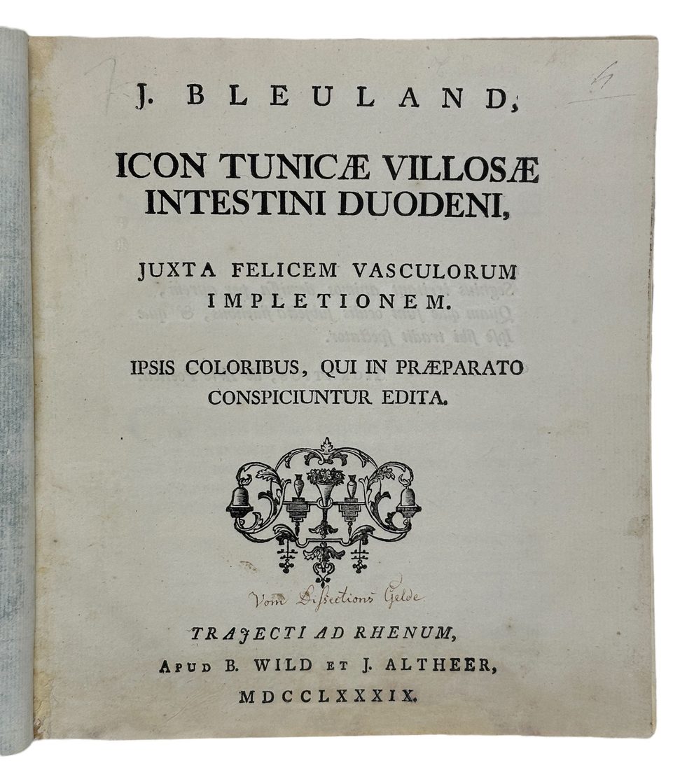
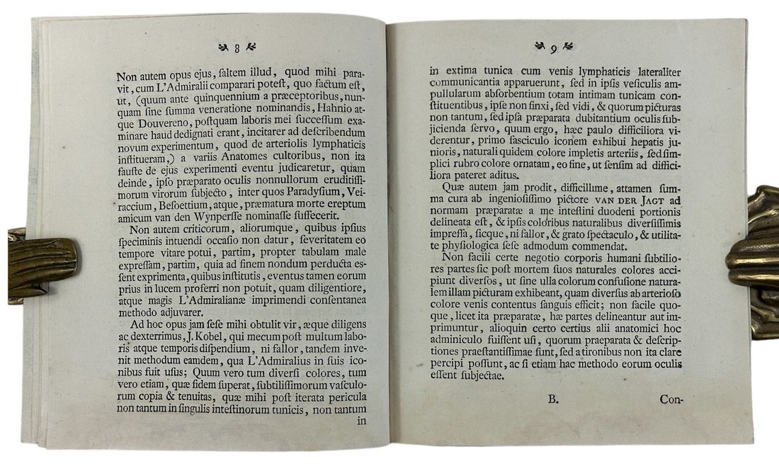
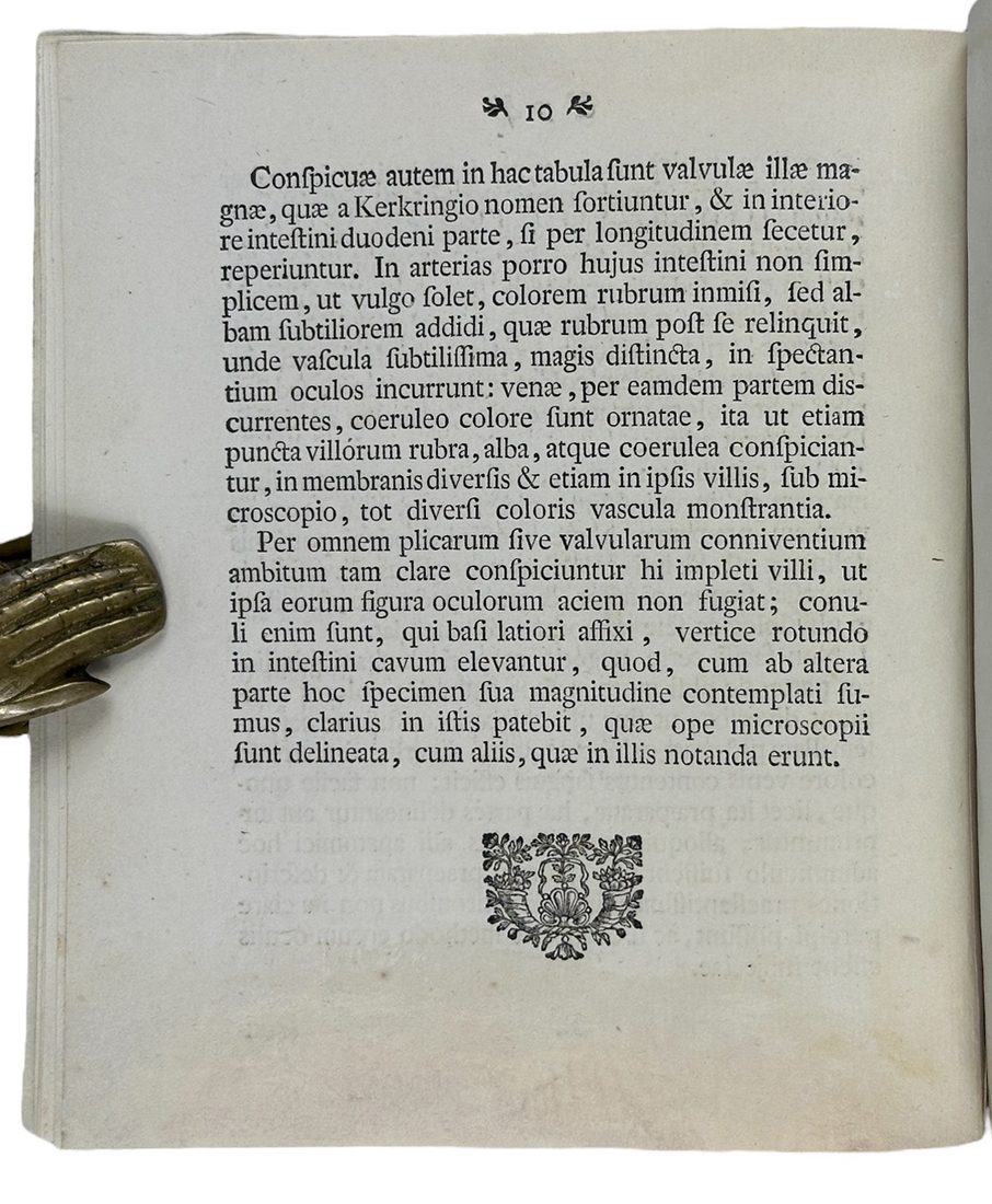
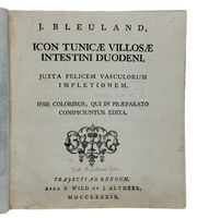
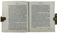
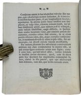
Bleuland, Jan. Icon tunicæ villosæ intestini duodeni, juxta felicem vasculorum impletionem. Ipsis coloribus, qui in præparato conspiciuntur edita. Trajecti ad Rhenum [Utrecht]: apud B. Wild et J. Altheer, 1789.
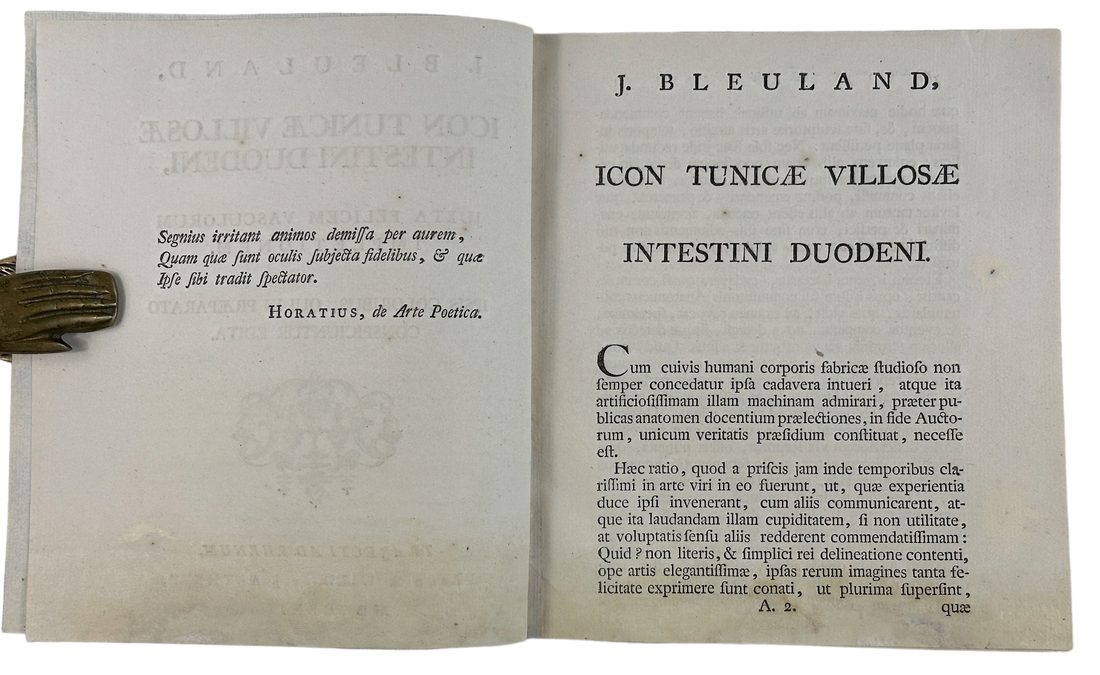
4to (21 x 19 cm), later paper wrapper, pp. 10, [2-last leaf blank missing] signature A⁴ B² (B2 blank, missing)
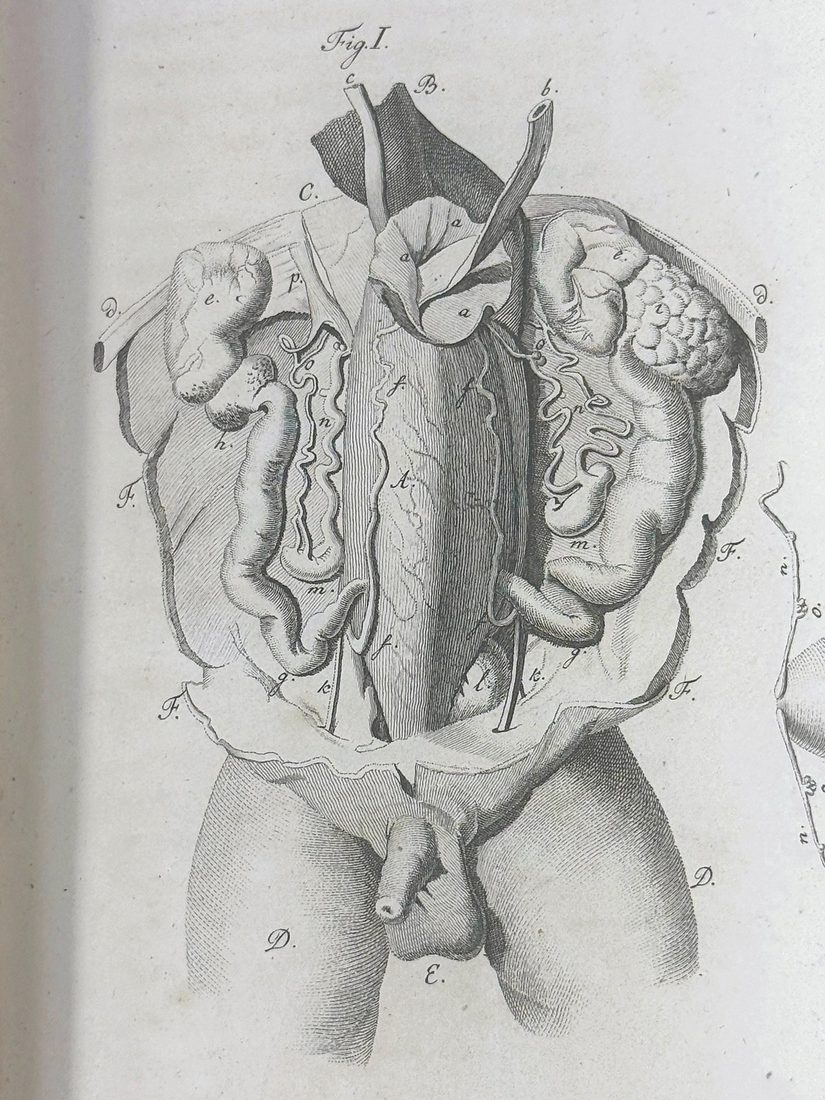
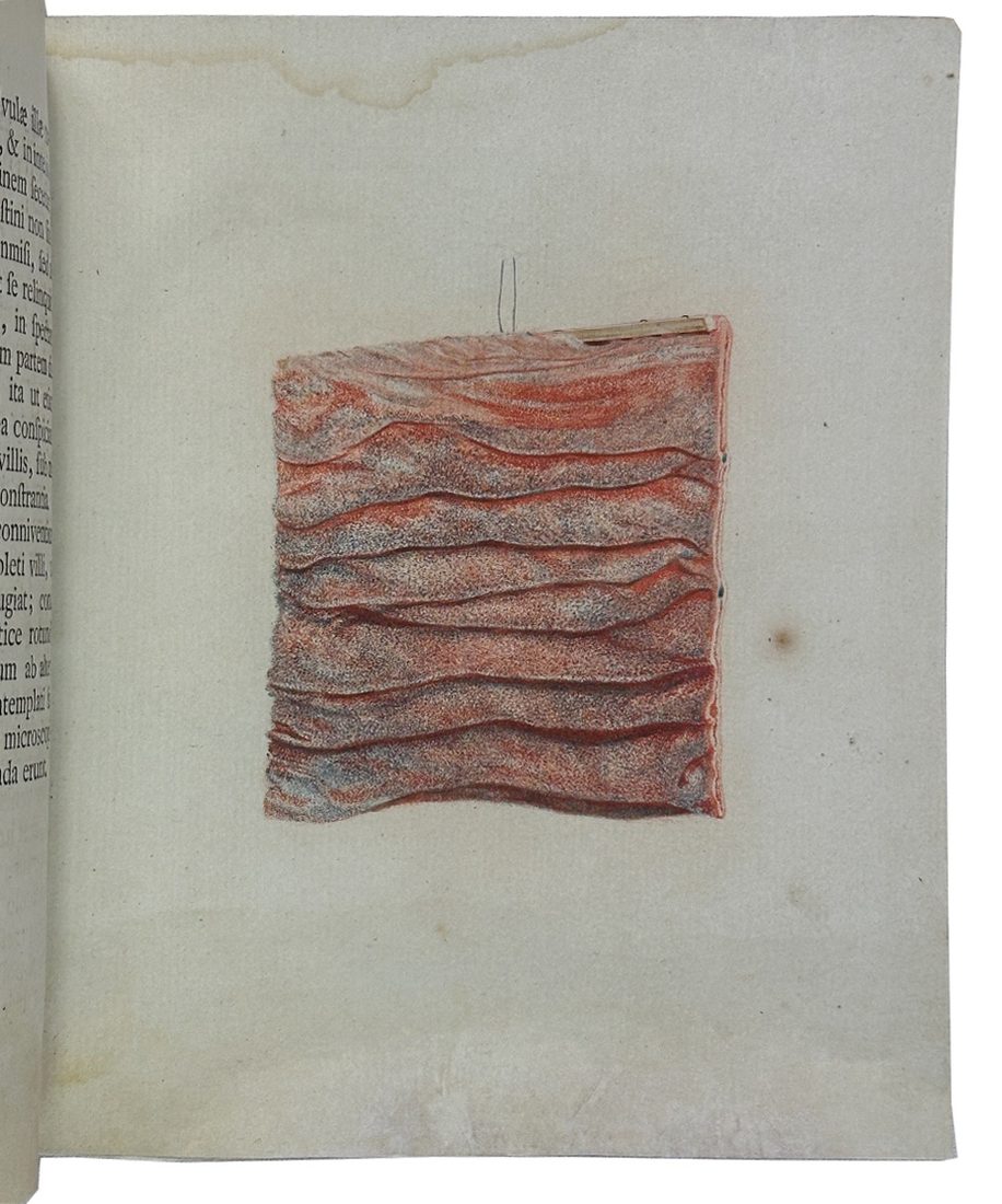
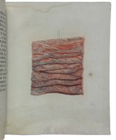
One coloured plate representing a sample of duodenal tissue
Jan Bleuland (1756-1838) was a renowned physician, professor of Anatomy, Physiology, Obstetrics and rector of Utrecht University.
He became famous for perfecting a technique of injecting fixatives and dyes into the soft tissues of the human body to reveal previously invisible anatomical details. The printed illustrations resulting from his technique are unique in their precision in depicting microscopic structures.
Among Bleuland's greatest achievements was his ability to visualize vascular systems, making even the smallest blood vessels perfectly visible, while preserving the true colours and textures of the tissues.
The ability to produce detailed and realistic tissue images, developed and improved by Bleuland, together with the work of his colleague, the Scottish pathologist Matthew Baillie (1761–1823), gave a significant boost to innovations in anatomical pathology.
During his distinguished career, Bleuland produced more than two thousand anatomical preparations, and his vast collection of specimens is now preserved at Utrecht Medical University.
“Bleuland focused on the soft parts [of human body], which involved a much wider spectrum of organs and diseases and posed a different set of problems to technicians in the preparation of the specimens” (Meli, p.211, op. cit.)
“During his career Bleuland published a number of illustrated anatomical works printed in color, showing a special interest in the fine structure of healthy and diseased states of the organs and tunics of the digestive tract. Bleuland made a collection of over two and a half thousand anatomical preparations, with the assistance of Petrus Koning, whom he had trained as his attendant and as a surgeon from 1800” (Meli, op.cit.)
Conditions: Small restoration at lower margins; overall a good copy.
Provenance: Ownership signature at title page
References: Domenico Bertoloni Meli, The Rise of Pathological Illustrations: Baillie, Bleuland, and Their Collections, Bulletin of the History of Medicine, Volume 89, Number 2, Summer 2015, pp. 209-242 (Article); Florian Rodari, Anatomie de la couleur, 124; James G. Mundies’s Cabinet of Curiosite’s, February 2008; Blake p. 50; not in Waller or Wellcome.